|
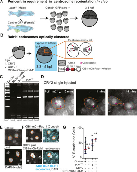
Pericentrin and Rab11 endosomes coordinate centrosome movement and number during mitotic exit.(A) Model depicting generation of pcnt+/− embryos with labeled centrosomes (centrin-GFP) to test the role of Pericentrin in centrosome reorientation and centrosome number in vivo. (B) Model depicting optogenetic clustering protocol for Rab11 endosomes. In short, embryos are injected with CRY2 and/or CIB1-mCh-Rab11 mRNAs, exposed to blue light 3.3–5 hpf causing a heterointeraction between CRY2 and CIB1, and imaged. Orange inset depicts cells imaged within the embryo. Pink circle, centrosome. (C) Ethidium bromide–stained acrylamide gel electrophoresis showing pcnt+/+ and pcnt+/− genotypes with ladder (lane 1), no template (lane 2), DNA extracted from control zebrafish (pcnt+/+, lane 3) and pcnt+/− embryos used in experimental analysis in Fig 6 (lane 4, 5, 6). (D) Time-lapse projections depicting a control dividing cell injected with CRY2 and PLK1-mCh (gray). Pink arrow, centrosome. Dashed lines, cell boundaries. Scale bar, 10 μm. (E, F) Representative images of fixed interphase zebrafish embryonic cells with a single nucleus or bi-nuclei labeled using DAPI in control, pcnt+/−, CIB1-mCh-Rab11 injected, and CRY2 plus CIB1-mCh-Rab11 injected embryos (gray, nuclei outlined with dashed orange line; E, F). Scale bar, 5 μm. (G) Percentage of embryos with cells containing binucleated cells was calculated for n > 30 cells per embryo across n > 3 embryos (G). One-way ANOVA with Dunnett’s multiple comparison with control, n.s. not significant, *P < 0.05 and **P < 0.01. n-values and statistical results detailed in Table S1.
|