|
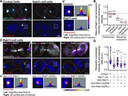
Rab11 GTP cycling mediates centrosome protein, Pericentrin, centrosome localization during pre-abscission.(A) Human (HeLa) pre-abscising cells expressing GFP-FIP3 (cyan) were fixed and immunostained for Pericentrin (magenta, top panel; fire LUT, bottom panel). Magnified insets (3×) shown on right (a’) with associated three-dimensional surface plot of intensity. Scale bar, 5 μm. (B) Box and whisker plot with mean (orange dashed line) depicting ratio of Rab11-null centrosome intensity over control centrosome intensity of cenexin, centrin, Pericentrin, FIP3, and transferrin receptor. Minimum and maximum values noted by boxed boundaries. n > 30 centrosomes per experiment across n = 3 experiments. One-way ANOVA with Dunnett’s multiple comparison to centrin, n.s. not significant, *P < 0.05 and ***P < 0.001. (C) GFP-FIP3 Rab11-null pre-abscising human (HeLa) cells ectopically expressing mCh-Rab11, -Rab11(S25N) or -Rab11(Q70L) (magenta, top panel) were fixed and immunolabeled for acetylated-tubulin (gray, top panel) and Pericentrin (fire LUT, bottom panel). Scale bar, 5 μm. (c’) 3× magnified centrosome inset (left), 3D fluorescent intensity surface plot of inset (right). (D) Scatter plot with median (orange dashed line) and quartiles (dark lines) depicting normalized Pericentrin intensities at centrosomes from pre-abscising human (HeLa) cells. One-way ANOVA with Dunnett’s multiple comparison to control, n.s. not significant and ****P < 0.0001. n > 38 centrosomes across n > 3 experiments. (B, D) n values and statistical results detailed in Table S1.
|