|
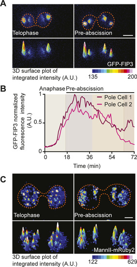
Mitotic centrosomes associate with Rab11 endosomes as they reorient towards the cytokinetic bridge.(A) Time-lapse of a pre-abscising human (HeLa) cell expressing GFP-FIP3 (16-color LUT). Bottom panels depict three-dimensional surface plot of the intensities portrayed in top panels. Dashed lines, cell boundaries. Scale bar, 5 μm. (A, B) Line graph depicting GFP-FIP3 normalized fluorescence intensity from anaphase exit to late abscission at the polar compartments from (A). Intensities of the brighter pole (pole 1), dark magenta and the less bright pole (pole 2), light pink. (C) Time-lapse of a pre-abscising human (HeLa) cell expressing MannII-mRuby2 (16-color LUT). Bottom panels depict three-dimensional surface plot of the intensities portrayed in top panels. Dashed lines, cell boundaries. Scale bar, 5 μm.
|