
Fig 2
- ID
- ZDB-FIG-211116-11
- Publication
- Bühler et al., 2021 - Histone deacetylase 1 controls cardiomyocyte proliferation during embryonic heart development and cardiac regeneration in zebrafish
- Other Figures
- All Figure Page
- Back to All Figure Page
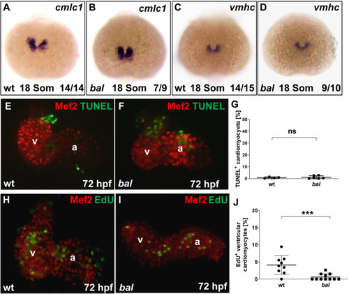
Early progenitor and chamber specification proceed normally in bal mutants.
(A-D) Whole-mount antisense RNA in situ hybridization (ISH) of cmlc1 and vmhc showed similar expression in bal mutants and wt siblings. (E, F) TUNEL staining (green) and IF for Mef2 (red) on dissected wt (E) and bal mutant hearts (F). (G) Quantification of TUNEL+ CMs revealed no alterations in apoptosis (wt 0.75 ± 0.70%, n = 4, bal 1.07 ± 1.28%, n = 6, p = 0.92), error bars indicate s.d., ns, not significant. (H, I) IF against Mef2 (red) and EdU (green) on dissected wt and bal hearts showed decreased numbers of EdU+ ventricular CMs. (J) Quantification revealed significantly decreased levels of EdU+ ventricular CMs in bal mutant hearts (wt 4.10 ± 2.73, n = 9, bal 0.61 ± 0.92, n = 11, p = 0.001). For significance testing Mann-Whitney-U test was applied, error bars indicate s.d., ***p < 0.001, ns, not significant. |
| Genes: | |
|---|---|
| Antibody: | |
| Fish: | |
| Anatomical Terms: | |
| Stage Range: | 14-19 somites to Protruding-mouth |
| Fish: | |
|---|---|
| Observed In: | |
| Stage: | Protruding-mouth |