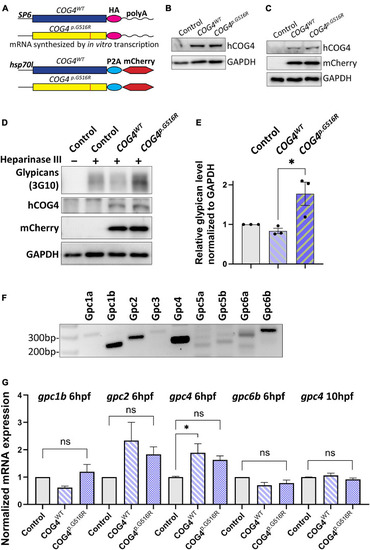
Expression of human COG4p.G516R in zebrafish increases the protein level of glypicans. (A–C) Expression of human COG4WT and COG4p.G516R in zebrafish after mRNA or DNA injection. (A) The scheme of COG4 constructs for in vitro transcription (top) and DNA injection (bottom). (B) Western blot at 24 hpf to detect the presence of COG4 after mRNA injection. (C) Western blot at 48 hpf to detect COG4 after DNA injection; heat shock was performed at 24 hpf for 2 h at 38°C. (D–G) Glypican analysis in zebrafish. (D) Western blotting of ΔHS-stub using 3G10 antibody following heparinase III digestion of control and embryos injected with COG4WT or COG4p.G516R mRNA at 3 dpf. (E) Quantitation assay of glypican band density in (D) and two more replicates. The data are presented as mean ± SEM. An unpaired two-tailed t-test was used. ∗p < 0.05. (F) mRNA expression of glypican genes by RT-PCR using cDNA from control embryos at 6 hpf. (G) qPCR analyses of highly expressed glypicans in control and zebrafish embryos injected with human COG4WT or COG4p.G516R mRNA. The relative glypican level was normalized to β-actin. The graphs represent the 2–ΔΔCt values. One-way ANOVA with Tukey’s multiple comparison tests was applied. ns, not significant; ∗p < 0.05. Experiments were performed in triplicates with similar results.
|
