FIGURE

FIGURE 1

ID
ZDB-FIG-211004-15
Publication
Xia et al., 2021 - A Dominant Heterozygous Mutation in COG4 Causes Saul-Wilson Syndrome, a Primordial Dwarfism, and Disrupts Zebrafish Development via Wnt Signaling
Other Figures
All Figure Page
Back to All Figure Page
FIGURE 1

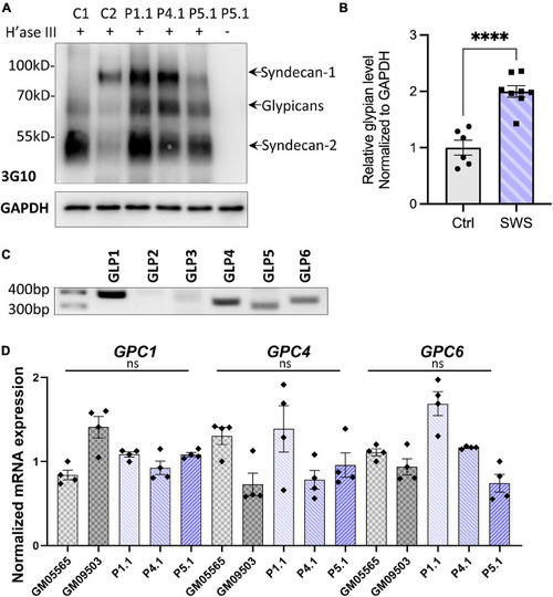
SWS-derived fibroblasts show altered HSPGs and glypicans after heparinase III (H’ase III) digestion. (A) Western blotting of ΔHS-stub using 3G10 antibody following heparinase III digestion of three SWS-derived fibroblasts and two control fibroblasts. C1 and C2 are control fibroblasts. C1, GM08429; C2, GM08680. P1.1, P4.1, and P5.1 are SWS-derived fibroblasts. (B) Quantitation assay of glypican band density in (A) and two other replicates. The data are presented as mean ± SEM. An unpaired two-tailed t-test was used. *⁣*⁣**p < 0.0001. (C) Agarose gel of six human glypicans after reverse-transcript PCR. (D) qPCR of three dominant glypicans in SWS-derived fibroblasts. The relative glypican level was normalized to GAPDH. The graphs represent the 2–ΔΔCt values. Experiments were performed in triplicates with similar results.

Expression Data

Expression Detail
Antibody Labeling
Phenotype Data

Phenotype Detail
Acknowledgments
This image is the copyrighted work of the attributed author or publisher, and ZFIN has permission only to display this image to its users. Additional permissions should be obtained from the applicable author or publisher of the image. Full text @ Front Cell Dev Biol