|
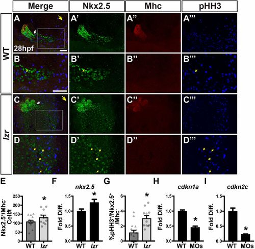
lzr mutants have an increase in proliferating SHFPs. (A-D‴) IHC of Nkx2.5, Mhc and pHH3 in WT and lzr mutant embryos at 28 hpf. B and D are higher magnification images of the boxed areas in A and C, respectively. Large yellow arrows in A and C indicate direction of the arterial pole of the heart. White arrows indicate the border between Nkx2.5+/Mhc+ and Nkx2.5+/Mhc− cells. Small yellow arrows in B, B′, B‴, D, D′ and D‴ denote cells co-expressing Nkx2.5 and pHH3. Scale bars: 50 µm (in A for A-A‴,C-C‴; in B for B-B‴,D-D‴). (E) Number of Nkx2.5+/Mhc− cells at 28 hpf in WT (n=16) and lzr (n=11) embryos. (F) RT-qPCR for nkx2.5 at 36 hpf in WT (n=4) and lzr mutant (n=4) embryos. (G) Percentage of Nkx2.5+/Mhc− cells co-expressing pHH3. WT (n=16), lzr (n=11). (H,I) RT-qPCR for cdkn1a and cdkn2c from sorted nkx2.5:ZsYellow+ cells isolated at 28 hpf (n=3). Error bars indicate s.e.m. *P<0.05.
|