
Fig. 8
- ID
- ZDB-FIG-200416-11
- Publication
- Holowiecki et al., 2020 - Pbx4 limits heart size and fosters arch artery formation through partitioning second heart field progenitors and restricting proliferation
- Other Figures
- All Figure Page
- Back to All Figure Page
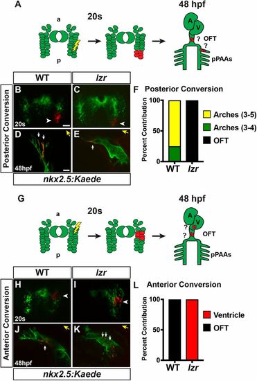
Posterior nkx2.5+ progenitors contribute to the OFT and ventricle in lzr mutants. (A,G) Approaches for photoconversion and analysis of posterior and anterior nkx2.5+ cells. At the 20 s stage, small clusters of nkx2.5:Kaede cells were converted from green to red. At 48 hpf, they were analyzed for contributions to the ventricle, OFT, and pPAAs. a, anterior; A, atrium; p, posterior; V, ventricle. (B,C,H,I) Confocal images of photoconverted nkx2.5:Kaede clusters (red; white arrowheads) from transgenic WT and lzr mutant embryos at the 20 s stage. Views are dorsal with anterior up. (D,E,J,K) Confocal images of the photoconverted cells from B, C, H and I at 48 hpf in the pPAA, OFT and ventricle. White arrows in D indicate photoconverted Kaede+ cells (red) in PAAs 3 and 4. Views are lateral with anterior right. White arrows in E and J indicate photoconverted Kaede+ cells (red) within the OFTs. White arrows in K indicate photoconverted Kaede+ cells within the ventricle. Yellow arrows indicate direction of the OFT. Scale bars: 50 µm. (F) Percentage of cells converted in the posterior that labeled pPAAs and OFT. WT (n=8), lzr mutants (n=3). (L) Percentage of cells converted in the anterior-medial region that labeled the ventricular CMs and OFT. WT (n=9), lzr (n=3). |