|
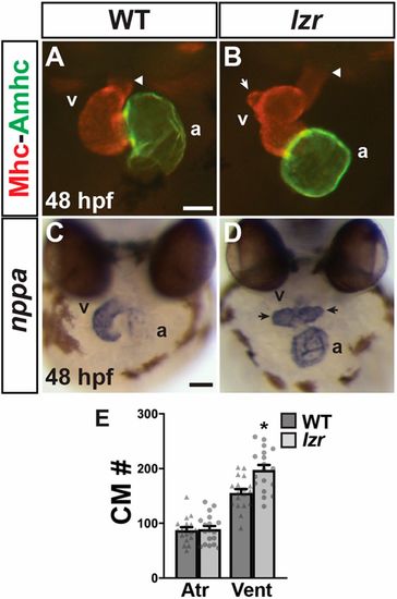
lzr mutants have an increase in ventricular CMs. (A,B) Hearts of WT and lzr embryos at 48 hpf. IHC for Sarcomeric myosin heavy chain (Mhc, red) and Atrial myosin heavy chain (Amhc, green). Images are frontal views. Arrowheads indicate the arterial pole of the heart. White arrow indicates a protrusion from the elongated OFT in a heart from a lzr embryo. Scale bar: 50 µm. (C,D) ISH for natriuretic peptide A (nppa). Images are frontal views. Arrows indicate ventricular protrusions. At least 30 embryos were examined for each condition. Scale bar: 100 µm. a, atrium; v, ventricle. (E) Number of CMs in WT (n=18) and lzr mutant (n=18) hearts with the myl7:DsRed2-NLS transgene. Atr, atrium; Vent, ventricle. Error bars indicate s.e.m. *P<0.05.
|