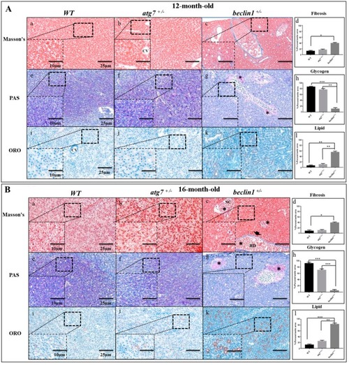
Defects of hepatic energy metabolism in male Beclin1+/− zebrafish. (A) Histological assessment of hepatocytes in 12-month-old male zebrafish. (a–d) Representative photomicrographs of Masson’s trichrome staining and the quantification of fibrosis. (e–h) Representative photomicrographs of Periodic Acid-Schiff (PAS) staining and glycogen content showing that glycogen packed in the hepatocytes of wild type (WT) and atg7+/− and lacked in beclin1+/− zebrafish; notice the beginning of hepatocellular malignancy (black star) around bile ducts. (i–l) Oil Red O (ORO) staining and lipid quantification revealing the alternations in fat droplet formation in beclin1+/− hepatocytes. (B) Histological assessment of hepatocytes in 16-month-old male zebrafish. (a–d) Representative photomicrographs Masson’s trichrome staining and fibrosis quantification with few fibrils lining the bile duct (arrowhead) and tumor-like cells (black stars) in beclin1+/−. (e–h) Representative photomicrographs of PAS staining with sufficient glycogen in WT and atg7+/− and sever glycogen reduction in beclin1+/−, notice the necrotic development around bile ducts (black stars). (i–l) ORO stain and lipid quantification showing hepatocytes hypertrophy with lipid accumulation and fatty clouds in beclin1+/− zebrafish. Scale magnification is shown in pictures and data are expressed as mean ± SD. * p < 0.05, ** p < 0.01, and *** p < 0.001. CV: Central Vein; BD: Bile Duct; NC: Necrosis.
|
