|
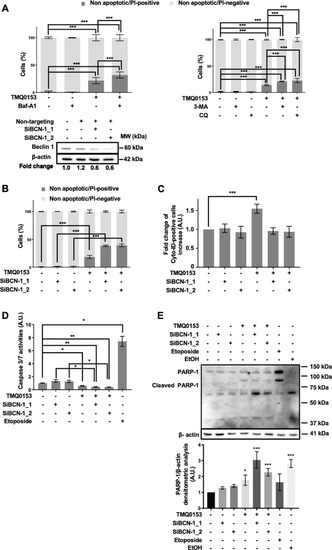
Inhibition of autophagy increases TMQ0153-induced necroptosis.a Effect of autophagy inhibitors (40 nM baf-A1, 10 mM, 3-methyladenine (3-MA) and 75 μM chloroquine (CQ)) on death of K562 cells treated with 30 µM of TMQ0153 assessed by nuclear morphology analysis after 8 h of treatment. b-e K562 cells were transfected with specific small interfering (si)RNAs against beclin 1 [(SiBCN-1), 5 nM SiBCN-1_1 and 10 nM SiBCN-1_2] for 24 h. b Upper panel: effect of siRNA on beclin 1 protein expression level. After quantification of the bands, beclin 1 levels were normalized to β-actin. Lower panel: transfected cells were treated with 30 μM of TMQ0153 and a nuclear morphology analysis was carried out after 8 h of treatment. c-e Effect of siRNA on TMQ0153-induced autophagy quantified by flow cytometry after Cyto-ID staining (c); on caspase-3/7 activity (d), and PARP-1 cleavage using C2-10 antibody (e). Etoposide (Eto; 100 µM, 24 h) and EtOH (10 %, 2 h) were used as positive controls for apoptotic and necrotic PARP-1 cleavage (upper panel) and the corresponding densitometric analysis of necrotic cleavage (lower panel) is visualized. In western blot analyses, β-actin was used as a loading control. All pictures are representative of three independent experiments and data represent mean (±S.D.) of three independent experiments Statistical significance was assessed as *p < 0.05, **p < 0.01, ***p < 0.001 for the indicated comparisons. Two-way ANOVA (nuclear morphology analysis); post hoc; Sidak’s test. One-way ANOVA (Cyto-ID assay); post hoc; Dunnett’s test. One-way ANOVA (caspase 3/7 assay); post hoc; Tukey’s test. One-way ANOVA (western blot quantification); post hoc; Dunnett’s test.
|