|
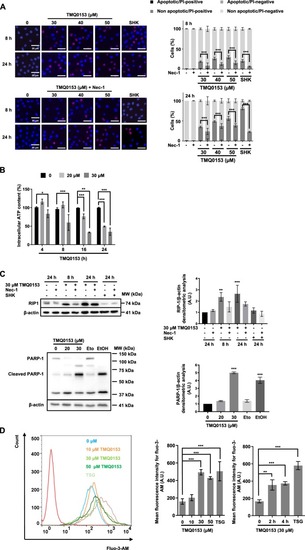
TMQ0153 induced a necrostatin-1-sensitive type of cell death in K562 cells.a K562 cells were incubated in presence or absence of 60 µM necrostatin (Nec)-1 for 1 h before a treatment with the indicated concentrations of TMQ0153. Shikonin (SHK; 5 µM) was used as a positive control for necrosis induction. a Nuclear morphology analyses by fluorescence microscopy following Hoechst/ propidium iodide (PI) staining after 8 and 24 h of treatment. Pictures representative of three independent experiments (left panels) and the corresponding quantification (right panels). b Measurement of intracellular ATP levels at the indicated concentrations and time points. c Receptor-interacting protein kinase (RIP)1 protein level and PARP-1 cleavage (left panel) were determined by western blotting using C2-10 antibody and the corresponding densitometric analysis (right panel). Shikonin (SHK; 5 µM, 24 h) and necrostatin (Nec)−1 (60 µM, 1 h) were used as a positive control and inhibitor for RIP1. Etoposide (Eto; 100 µM, 24 h) and ethanol (EtOH; 10 %, 2 h) were used as positive controls for apoptotic and necrotic PARP-1 cleavage, respectively. d Cytosolic Ca2+ levels were measured using Fluo-3-AM after 24 h (left panel) and time-dependently measured Ca2+ levels at 30 μM (right panel). Thapsigargin (TSG; 300 nM, 24 h) was used as a positive control for intracellular Ca2+ accumulation. β–actin was used as loading control. All pictures are representative of three independent experiments and all graphs represent the mean (±S.D.) of three independent experiments. Statistical significance was assessed as *p < 0.05, **p < 0.01, ***p < 0.001 for the indicated comparisons. Two-way ANOVA (microscopy analysis, cell viability); post hoc: Sidak’s test. Two-way ANOVA (Cell Titer Glo assay); post hoc; Tukey’s test. One-way ANOVA (intracellular Ca2+ assay); post hoc; Dunnett’s test. One-way ANOVA (western blot quantification); post hoc; Dunnett’s test.
|