
Fig. S5
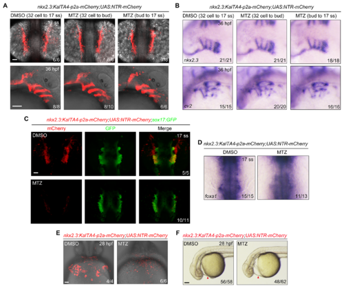
NTR-mediated ablation of nkx2.3+ progenitors impairs pericardium development but not endoderm formation.(A-B) Tg(nkx2.3:KalTA4-p2a-mCherry;UAS:NTR-mCherry) embryos were treated with 50 mM MTZ during different time intervals and then harvested at the indicated developmental stages for in vivo confocal imaging (A) and in situ hybridization (B). Scale bars, 50 μm. (C-D) Depletion of nkx2.3+ progenitors has no obvious effect on endoderm formation. Tg(nkx2.3:KalTA4-p2a-mCherry;UAS:NTR-mCherry;sox17:GFP) or Tg(nkx2.3:KalTA4-p2a-mCherry;UAS:NTR-mCherry) embryos were treated with 50 mM MTZ from the 32-cell stage to the 17-somite stage. Then these embryos were subjected to in vivo confocal imaging (C) and in situ hybridizations (D) at the 17-somite stage. In panel D, embryos are viewed from the dorsal aspect, and the white dotted lines indicate the region of the pericardium. Scale bars, 50 μm. (E-F) Depletion of nkx2.3+ progenitors leads to obvious pericardial edema. Tg(nkx2.3:KalTA4-p2a-mCherry;UAS:NTR-mCherry) embryos were treated with 50 mM MTZ from the 32-cell stage to the 17-somite stage, and then these embryos were harvested at 28 hpf for in vivo confocal imaging (E, ventral views, anterior to the top; Scale bar, 50 μm). Their morphological defects were shown in (F, lateral views with anterior to the left; Scale bar, 100 μm). Red Arrowheads indicate the pericardium. |