|
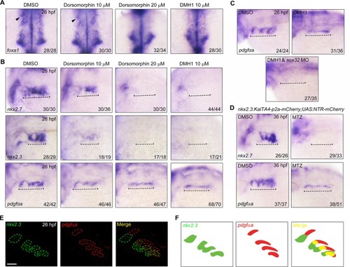
BMP signal inhibition results in a severe loss of <italic>nkx2</italic>.<italic>3</italic><sup>+</sup> pouch epithelium.(A-B) In situ hybridization analysis of the expression of foxa1 (A), nkx2.7, nkx2.3, and pdgfαa (B) at 26 hpf in embryos treated with different dose of BMP inhibitors from bud stage to the 17-somite stage. Black arrowheads in (A) indicate the first pouch. Black dotted lines in (B) highlight the pharyngeal region. (C) The expression of pdgfαa in DMH1-treated embryos injected with or without sox32 MO. Note that when DMH1-treated embryos were injected with 8 ng sox32 MO, the expression of pdgfαa was abolished. (D) The expression of nkx2.7 and pdgfαa in nkx2.3+ pouch progenitor-depleted embryos. Tg(nkx2.3:KalTA4-p2a-mCherry;UAS:NTR-mCherry) embryos were treated with 50 mM MTZ from the 32-cell stage to the 17-somite stage, and then harvested for in situ hybridization at 36 hpf. (E-F) Double-fluorescence in situ hybridizations for nkx2.3 and pdgfαa at 26 hpf, In situ hybridization analysis of pdgfαa at 26 hpf. Note that nkx2.3 (green) and pdgfαa (red) display nested expression patterns along the dorsal-ventral axis of the pouch epithelium. Schematic expression of nkx2.3 and pdgfαa was shown in (F). Scale bar, 50 μm.
|