|
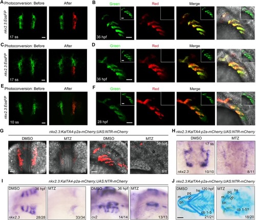
<italic>nkx2</italic>.<italic>3</italic><sup>+</sup> progenitors give rise to pharyngeal pouches.(A-B) Tg(nkx2.3:EosFP) embryos at the 17-somite stage before (green) and after (red) photoconversion in the right-side nkx2.3+ cluster (A). At 36 hpf, embryos were imaged in the green and red channels (B). Cells in the right-side nkx2.3+ cluster remained un-photoconverted as an internal control and their derivatives were imaged and shown in the inset. Scale bars, 50 μm. (C-D) The posterior part of the right-side nkx2.3+ cluster was photoconverted at the 17-somite stage (C). Images of the pharyngeal pouches in the same embryos at 36 hpf are shown in (D). Scale bars, 50 μm. (E-F) Tg(nkx2.3:EosFP) embryos were photoconverted in the right-side nkx2.3+ cluster at the 10-somite stage (E), and then these embryos were imaged in the red and green channels (inset) at 28 hpf (F). Scale bars, 50 μm. (G-J) Tg(nkx2.3:KalTA4-p2a-mCherry;UAS:NTR-mCherry) embryos were treated with 50 mM MTZ from the 32-cell stage to the 17-somite stage. Subsequently, these embryos were harvested at the indicated developmental stages for in vivo confocal imaging (G), in situ hybridization (H-I) and Alcian Blue staining (J). m, Meckel’s; pq, palatoquadrate; hs, hyosymplectic; bh, basihyal; ch, ceratohyal; cb, ceratobranchial. Scale bars, in panel G, 50 μm; in panel J, 100 μm.
|