|
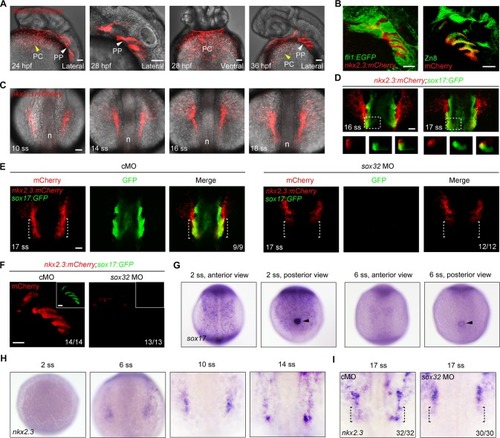
<italic>nkx2</italic>.<italic>3</italic> is expressed in the lateral pharyngeal endoderm during early somite stages.(A) Tg(nkx2.3:mCherry) embryos at 24, 28 and 36 hpf exhibiting fluorescence in the pericardium (yellow arrowhead) and pharyngeal pouches (white arrowhead). PC, pericardium; PP, pharyngeal pouch. Scale bars, 50 μm. (B) mCherry-positive cells (red) were located between EGFP-labeled cranial neural crest cells (green) in Tg(nkx2.3:mCherry;fli1:EGFP) embryos (left panel), and co-localized with Zn8 labeled pharyngeal pouch cells (green) in Tg(nkx2.3:mCherry) embryos (right panel). Scale bars, 50 μm. (C) mCherry fluorescence in Tg(nkx2.3:mCherry) embryos from the 10- to 18-somite stages. Embryos were dorsal views with anterior to the top. n, notochord; ss, somite stage. Scale bar, 50 μm. (D) Tg(nkx2.3:mCherry;sox17:GFP) embryos with mCherry-positive cells (red) and GFP-labeled endodermal cells (green) at the 16- and 17-somite stages. The lower panels are optical transverse sections (XZ) taken at the level of white lines in their respective upper panels. Scale bar, 50 μm. (E-F) mCherry (red) and GFP (green) fluorescence in Tg(nkx2.3:mCherry;sox17:GFP) embryos injected with 8 ng control MO (cMO) or 8 ng sox32 MO at the 17-somite stage (E) and 36 hpf (F). In panel F, the GFP fluorescence was shown in the inset. The ratios of affected embryos are indicated. White dotted lines highlight the lateral pharyngeal endoderm. Scale bars, 50 μm. (G-H) In situ hybridization of sox17 (G) and nkx2.3 (H) expression in wild-type embryos at indicated developmental stages. Black arrowheads in (G) indicate the KVs. (I) Alteration of nkx2.3 expression pattern in 8 ng sox32 MO injected embryos at the 17-somite stage. Black dotted lines show the lateral pharyngeal endoderm.
|