|
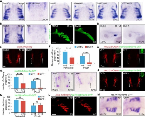
BMP signal inactivation leads to a decrease of pouch progenitors.(A-B) Wild-type embryos were exposed to different inhibitors from bud stage to the 17-somite stage and harvested at 36 hpf for in situ hybridizations with nkx2.3 probe. Dorsal views, anterior to the top. Note that embryos treated with 20 μM Dorsomorphin (A) or 10 μM DMH1 (B) showed a clear reduction of nkx2.3 expression in pharyngeal pouches. (C) Live confocal images of malformed pharyngeal pouches in 10 μM DMH1 treated Tg(sox17:GFP) embryos at 36 hpf. Scale bar, 50 μm. (D) In situ hybridization of thymus marker ccl25a in DMH1 treated embryos at 48 hpf. Lateral views, anterior to the left. (E-F) Representative confocal sections showing mCherry+ progenitors in embryos treated with or without 10 μM DMH1 from bud stage to the 17-somite stage (E). The lateral pharyngeal endoderm is indicated by white dotted lines. Scale bar, 50 μm. Quantification of the numbers of pericardial and pouch progenitors positive for mCherry in DMSO and DMH1 conditions was shown in (F). The group values are expressed as mean±s.d. Student’s t-test, ****P<0.0001. (G-H) mCherry fluorescence in 9 hpf-heat shocked Tg(nkx2.3:mCherry;hsp70l:dnBmpr1a-GFP) embryos at the 17-somite stage (G). Scale bar, 50 μm. The numbers of mCherry+ progenitors were quantified from heat shocked-embryos with or without GFP expression (H). Student’s t-test, ****P<0.0001. (I) The expression of nkx2.3 in embryos treated with 10 μM DMH1 from bud stage to the 17-somite stage. Black dotted lines indicate the region where the pouch progenitors are located. (J-M) Overactivation of BMP signaling has no effect on the formation of nkx2.3+ pouch progenitors and pouch epithelium. Tg(nkx2.3:mCherry;hsp70l:caBmpr1b-GFP) embryos were heat shocked at 9 hpf for 20 min, and then harvested at the indicated developmental stages for in vivo confocal imaging (J-L) and in situ hybridization (M). Scale bars, 50 μm. The number of mCherry+ progenitors was calculated from heat shocked-embryos in (J) and presented in (K). ns, non-significant.
|