
Fig. 7
- ID
- ZDB-FIG-191007-9
- Publication
- Nicholas et al., 2019 - Temporal characterization of optic fissure basement membrane composition suggests nidogen may be an initial target of remodeling
- Other Figures
- All Figure Page
- Back to All Figure Page
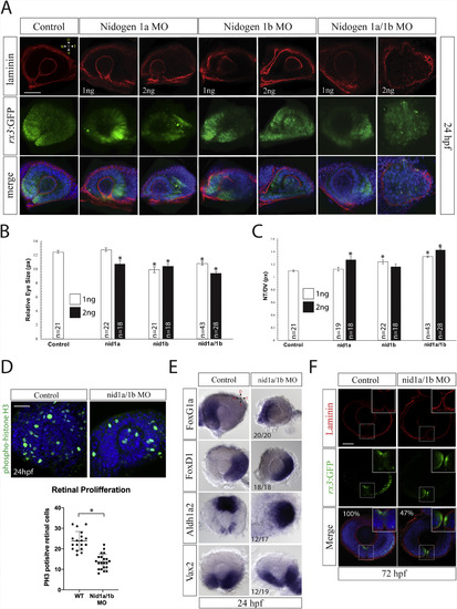
Nidogen is essential for accurate retinal morphogenesis. A)Retinal phenotypes in 24hpf nid1 morphant rx3:GFP (green) expressing embryos were characterized using laminin IHC (red), and DAPI (blue). Representative single confocal sections are depicted. Retinal shape is moderately disrupted at low morpholinoconcentrations (1 ng) with most severe effects observed in double morphant embryos (2 ng). Laminin deposition appears to be mildly affected upon co-injection of nid1a and 1b MO at 2 ng 40–50 embryos were examined for each treatment. Scale bar = 50 μm. B) Quantification of relative retinal size observed in 24hpf morphant embryos. Significantly smaller eyes were observed upon injection of either nid1a or nid1b morpholinos, with the smallest resulting from co-injection of nid1a and 1b morpholinos. Student T-tests were conducted between control and all treatment groups for each parameter measured. * = p < .005. C) Quantification of retinal shape in 24hpf nidogen morphant embryos. Injection of nid1a or nid1b results in an extended NT axis, with co-injection having a synergistic effect. Student T-tests were conducted between control and all treatment groups for each parameter measured. * = p < .05. D) Phospho-histoneH3 (PH3) staining (green) at 24hpf in control and nid1a/1b co-injected embryos. 3D reconstructions are displayed. PH3+ cell counts from individual embryos analyzed are graphed. DAPI is shown in blue. WT n = 19, Nid1a/1b MO n = 23. *p = .0001, Student T-test. Scale bar = 50 μm. E)WISH for temporal, foxG1a, nasal, foxD1, ventral, vax2 and dorsal, aldh1a2 in nid1a/nid1b morphant retinas at 24hpf. WT and morphant embryo eyes were dissected, mounted and imaged. Nid1a/1b morphant eyes show no significant effects on nasal/temporal patterning, while ventral domains appear shifted temporally and dorsal regions expanded. F) Examination of optic fissure fusion at 72hpf via laminin (red) IHC in rx3:GFP (green) expressing morphant embryos. Inset (dashed square) displays high magnification of the optic fissure. Single confocal planes of the central-distal region of the fissure are depicted. Results reveal the persistence of laminin signal in 47% (n = 41 control, n = 27 nid1a/1b MO, p = .0001) of 72hpf morphant fissures, a time when control embryos have already completed fusion and removed laminin. Scale bar = 50 μm. |
| Genes: | |
|---|---|
| Antibodies: | |
| Fish: | |
| Knockdown Reagents: | |
| Anatomical Terms: | |
| Stage Range: | Prim-5 to Protruding-mouth |
| Fish: | |
|---|---|
| Knockdown Reagents: | |
| Observed In: | |
| Stage: | Prim-5 |
Reprinted from Developmental Biology, 452(1), Nicholas, C., Weaver, M., Piedade, W.P., Vocking, O., Famulski, J.K., Temporal characterization of optic fissure basement membrane composition suggests nidogen may be an initial target of remodeling, 43-54, Copyright (2019) with permission from Elsevier. Full text @ Dev. Biol.