
Fig. S3
- ID
- ZDB-FIG-180622-22
- Publication
- Di Donato et al., 2018 - An Attractive Reelin Gradient Establishes Synaptic Lamination in the Vertebrate Visual System
- Other Figures
- All Figure Page
- Back to All Figure Page
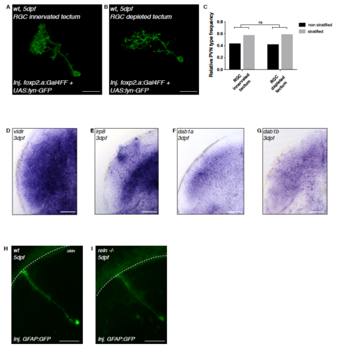
PVN stratification in eyeless larvae and Reelin signaling pathway member expressions in PVNs. Related to Figure 2. (A) Confocal image of a transiently GFP-labeled PVN in an RGC-innervated tectum at 5dpf shown from the side, parallel to the skin to highlight the PVNs’ stratified laminar morphology. (B) Confocal image of a transiently GFP-labeled PVN in an RGC-depleted tectum at 5dpf shown from the side, parallel to the skin to highlight the PVNs’ stratified laminar morphology. (C) Frequency distribution of non-stratified PVNs and stratified PVNs without lamina targeting mistakes at 5-6dpf in RGC-depleted tecta (n=7 larvae) and the tectum of larvae of which the corresponding eye was removed at 2 dpf (n=12 larvae). We could not observe any difference between the two groups (k*2-chi-square test after Brandt-Snedecor, p = 0.9596) thus suggesting that PVN lamination defects seen in reln -/-, vldlr -/-,/sup> and dab1a -/- are not a consequence of prior RGC mis-targeting but are better explained by PVNs using Reelin signaling as guidance signal to identify single target lamina. (D) Horizontal cross-section of a 3 dpf zebrafish tectum showing strong mRNA expression of the Reelin receptor vldlr in PVNs. (E) Horizontal cross-section of a 3 dpf zebrafish tectum showing strong mRNA expression of the Reelin receptor lrp8 in PVNs. (F) Horizontal cross-section of a 3 dpf zebrafish tectum showing mRNA expression of dab1a, an intracellular transducer downstream of Reelin receptors, in PVNs. (G) Horizontal cross-section of a 3 dpf zebrafish tectum showing mRNA expression of dab1b, a paralog of dab1a, in PVNs. (H-I) Confocal reconstruction of radial glia cells transiently labeled with the GFAP:GFP construct. Radial glia span the entire depth of the larval zebrafish tectum, from the ventricular region to the surface of the tectal neuropil (white dashed line) in both wild-type (n=6) and reln -/- (n=6) larvae at 5 dpf, suggesting that radial fibers are not contributing to layer-specific guidance of RGC axons by Reelin. |
| Genes: | |
|---|---|
| Fish: | |
| Anatomical Term: | |
| Stage: | Protruding-mouth |