
Fig. 5
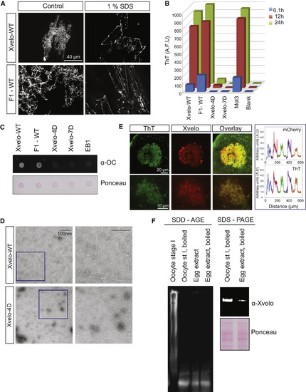
Xvelo Shows Amyloid-like Features In Vivo and In Vitro (A) SDS was added to Xvelo and F1-WT-GFP networks to a final 1% concentration, and the reactions were incubated at room temperature for 15 min. The resulting mixtures were squashed under a coverslip and imaged by a spinning-disc confocal microscope. (B) A final concentration of 5 μM of Thioflavin T was added to the wild-type, F1, and mutant network reactions at the indicated time points. Yeast prion Mot3 was used as a positive control, whereas blank was only buffer and ThT. ThT fluorescence was measured (a.u.) by a fluorescence plate reader. (C) 1 μg RFP-tagged wild-type, F1, and mutant recombinant proteins were dot-blotted on a nitrocellulose membrane and assayed for reactivity with α-amyloid fibril OC. EB1-RFP was used as a negative control. (D) Negative stain electron microscopy images of the untagged Xvelo-WT and Xvelo-4D self-assembly reactions (scale bars, 100 nm). (E) Stage I oocytes were injected with mRNA coding for Xvelo-mCherry and incubated overnight. The oocytes were incubated in 10 μM ThT, washed twice, and imaged by confocal microscopy. Bottom: zoomed in images. Line scans showing the co-localization of Xvelo-mCherry and ThT stain from five Balbiani bodies were plotted. Each color represents the line scan of a different Balbiani body. We speculate that the outer rim Xvelo-mCherry signal belongs to the newly translated Xvelo-mCherry protein that has just started to form a new, immature matrix and does not yet stain with ThT. (F) SDD-AGE detects SDS-resistant Xvelo aggregates in vivo. Equal amounts of cytoplasmic extracts of stage I oocytes and mature eggs were loaded onto SDS-PAGE. A five times more amount of egg extracts was loaded for SDD-AGE gels to make Xvelo concentrations comparable between the oocyte and egg extract lanes. Xvelo was detected by an anti-Xvelo antibody. See also Figure S5. |
Reprinted from Cell, 166, Boke, E., Ruer, M., Wühr, M., Coughlin, M., Lemaitre, R., Gygi, S.P., Alberti, S., Drechsel, D., Hyman, A.A., Mitchison, T.J., Amyloid-like Self-Assembly of a Cellular Compartment, 637-50, Copyright (2016) with permission from Elsevier. Full text @ Cell