
Fig. 2
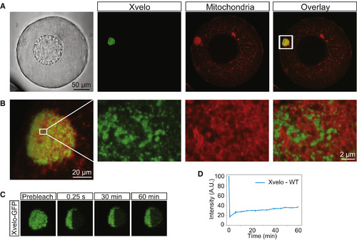
Xvelo Forms a Stable Matrix (A) mRNA encoding for Xvelo-GFP was microinjected into stage I oocytes. MitoTracker Deep Red was used to label mitochondria. Oocytes were imaged live with a laser scanning confocal microscope with a 40× water-immersion objective. (B) Magnification of the Balbiani body in (A). (C) Internal rearrangement of fluorescent Xvelo-GFP particles after half bleach over time. (D) The fluorescent recovery of the half-bleached Xvelo-GFP in the Balbiani body in (C) and two other biological replicates is shown by quantification of fluorescence in bleached region over time. Fluorescent intensity changes in the bleached region per pixel over time were plotted after it was normalized for photobleaching by using an unbleached neighboring area and background subtraction. See also Figure S2. |
Reprinted from Cell, 166, Boke, E., Ruer, M., Wühr, M., Coughlin, M., Lemaitre, R., Gygi, S.P., Alberti, S., Drechsel, D., Hyman, A.A., Mitchison, T.J., Amyloid-like Self-Assembly of a Cellular Compartment, 637-50, Copyright (2016) with permission from Elsevier. Full text @ Cell