
Fig. 1
- ID
- ZDB-FIG-150512-1
- Publication
- Burgoyne et al., 2015 - Regulation of melanosome number, shape and movement in the zebrafish retinal pigment epithelium by OA1 and PMEL
- Other Figures
- All Figure Page
- Back to All Figure Page
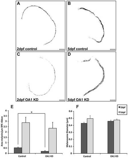
OA1 MOs reduce melanosome number at 2dpf without affecting melanosome size. (A–D) Contrast-enhanced images of zebrafish eye cross-sections highlighting only electron-dense melanin. Images are shown from (A,B) control and (C,D) OA1 MO-treated zebrafish. KD, knockdown. Scale bars: 20µm. (E) At 2dpf there is a significant reduction in melanin area between controls and OA1 MO-treated zebrafish. By 5dpf, the OA1 MO appears to be less effective, resulting in no significant difference in melanin area between controls and OA1 MO-treated animals. (F) The OA1 MO had no apparent effect on melanosome diameter at 2 and 5dpf. Results show the mean±s.e.m.; *P<0.05 (Student′s t-test). |
| Fish: | |
|---|---|
| Knockdown Reagent: | |
| Observed In: | |
| Stage Range: | Long-pec to Day 5 |