
FIGURE
Fig. 2
- ID
- ZDB-FIG-150512-2
- Publication
- Burgoyne et al., 2015 - Regulation of melanosome number, shape and movement in the zebrafish retinal pigment epithelium by OA1 and PMEL
- Other Figures
- All Figure Page
- Back to All Figure Page
Fig. 2
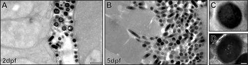
Intense biogenesis of mature melanosomes between 2 and 5dpf. Electron micrographs of zebrafish RPE show a large increase in melanosome number between 2dpf (A) and 5dpf (B). (C,D) High-magnification images of mature melanosomes at 2dpf. Some mature melanosomes have an appearance of ‘holes’ (D). Scale bars: 1µm (A,B), 100nm (C,D). |
Expression Data
Expression Detail
Antibody Labeling
Phenotype Data
Phenotype Detail
Acknowledgments
This image is the copyrighted work of the attributed author or publisher, and
ZFIN has permission only to display this image to its users.
Additional permissions should be obtained from the applicable author or publisher of the image.
Full text @ J. Cell Sci.