
Fig. S1
- ID
- ZDB-FIG-150331-43
- Publication
- Wakayama et al., 2015 - Cdc42 Mediates Bmp-Induced Sprouting Angiogenesis through Fmnl3-Driven Assembly of Endothelial Filopodia in Zebrafish
- Other Figures
- All Figure Page
- Back to All Figure Page
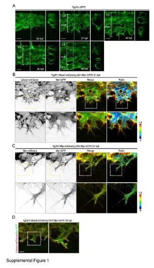
Visualization of F-actin in Endothelial Cells (ECs) during Caudal Vein Plexus (CVP) Formation. Related to Figure 1. A) Confocal z-stack images of the caudal regions of Tg(fli1:GFP) embryos at the developmental stages indicated at the right bottom corner of the images. The cross-sectional images of the areas indicated by dotted lines are shown in the panels to the right of the original images. CA, caudal artery; CV, caudal vein; CVP, caudal vein plexus. (B) 3D-rendered confocal microscopic images of the CVP of Tg(fli1:lifeact-mCherry);(fli1:Myr-GFP) embryo at 31 hpf. mCherry (Lifeact-mCherry) and GFP (Myr-GFP) images, their merged images (GFP, green; mCherry, red) and the mCherry/GFP ratio images shown in the intensity-modulated display (IMD) mode (Ratio) are shown as indicated at the top. The upper and lower limits of the ratio range are indicated on the right. The boxed areas are enlarged on the panels beneath the original images. (C) Confocal 3D images of the CVP of Tg(fli1:Myr-mCherry);(fli1:Myr-GFP) embryo at 31 hpf. mCherry (Myr-mCherry) and GFP (Myr-GFP) images, their merged images (GFP, green; mCherry, red) and the mCherry/GFP ratio images shown in the IMD mode (Ratio) are shown similar to B. (D) Confocal z-stack images of the CVP of Tg(fli1:Myr-mCherry);(fli1:Myr-GFP) embryo at 29 hpf. The merged images of mCherry (Lifeact-mCherry) and GFP (Myr-GFP) are shown. The boxed area is enlarged in the right panel. Scale bars: 50 µm (A) and 20 µm (D). |
| Genes: | |
|---|---|
| Fish: | |
| Anatomical Terms: | |
| Stage Range: | Prim-5 to Long-pec |
Reprinted from Developmental Cell, 32, Wakayama, Y., Fukuhara, S., Ando, K., Matsuda, M., Mochizuki, N., Cdc42 Mediates Bmp-Induced Sprouting Angiogenesis through Fmnl3-Driven Assembly of Endothelial Filopodia in Zebrafish, 109-22, Copyright (2015) with permission from Elsevier. Full text @ Dev. Cell