Fig. 7
|
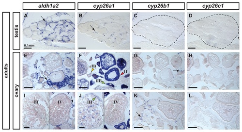
Expression patterns of aldh1a2, cyp26a1, cyp26b1 and cyp26c1 in adult gonads. In adult testes (n=3), aldh1a2 expression was detected in somatic cells surrounding cysts in a localization expected for Sertoli cells (A) and cyp26a1 expression was detected in a subset of cells in a localization expected for Leydig cells (B). The presence of germ cells expressing cyp26a1 could not be discarded. Expression of cyp26b1 (C) and cyp26c1 (D) was not detected in adult testes. In adult ovaries (n=2), aldh1a2 was detected in somatic cells but not in the oocytes (E,I). Expression of cyp26a1 was restricted to the ooplasm of oocytes and was not detected in somatic cells (F,J). In oocytes, cyp26a1 expression varied according to the stage of meiosis: it was barely detectable in early stage IB oocytes (eIB, yellow arrowhead in F, prior to the diplotene stage of meiosis), was up-regulated in late stage IB oocytes that entered meiotic arrest at diplotene stage (lIB, green arrowhead in F), was maintained in stage II (red arrowhead in F) and stage III oocytes (green arrowhead in J) and was not detected in stage IV oocytes (J) coinciding with the resumption of meiosis I. Expression of cyp26b1 was detected solely in a subset of cells in the somatic tissue surrounding the oocytes (arrows in G, K) and no expression of cyp26c1 was detected in the ovary (H,L). Scale bar: 0.1mm. |
| Genes: | |
|---|---|
| Fish: | |
| Anatomical Terms: | |
| Stage: | Adult |

