
Fig. 4
- ID
- ZDB-FIG-130907-4
- Publication
- Alunni et al., 2013 - Notch3 signaling gates cell cycle entry and limits neural stem cell amplification in the adult pallium
- Other Figures
- All Figure Page
- Back to All Figure Page
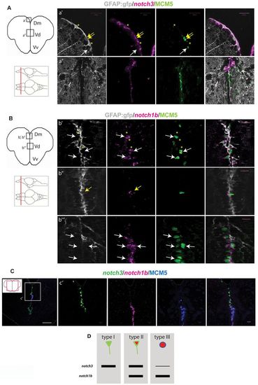
notch3 and notch1b are differentially expressed in adult pallial progenitors. (A) notch3 expression revealed by fluorescent in situ hybridization (magenta) on telencephalic cross-sections of a gfap:gfp brain, together with a double fluorescent immunostaining for GFP (RG, gray) and MCM5 (green). (a2) Dorsomedial (Dm) region of the pallium, notch3 expression is found in quiescent (type I) (yellow asterisks) and proliferating (type II) (yellow arrows) RG. Transcripts are absent from type III cells (white arrows). (a2) Pallial-subpallial junction, enriched in type III cells: notch3 expression is very low or absent. (B) notch1b expression (magenta) compared with GFAP-GFP (RG, gray) and MCM5 (green). (b2,b2) Dm; white and yellow arrows indicate notch1b-positive type II RG (b2) and type III progenitors (b2), respectively. Transcripts are absent from quiescent RG (b2, yellow asterisks). (b22) Pallial-subpallial junction: notch1b is strongly expressed in type III cells. (C) Double fluorescent in situ hybridization for notch3 (green) and notch1b (magenta) with immunostaining for MCM5 (blue). (c2) High magnification of the pallial-subpallial junction: notch3 is expressed by RG cells of Dm and notch1b by type III progenitors. (D) Graphic representation of notch3 and notch1b expression in the different progenitor cell types (according to März et al., 2010). In Dm, notch3 is expressed by 97% of type I cells (n=229 cells counted), 88% of type II cells (n=31 cells) and 2% of type III cells(n=23 cells); notch1b is expressed by 2.8% of type I cells (n=141 cells), 90% of type II cells (n=43 cells) and 85% of type III cells. Scale bars: white, 100 μm; magenta, 20 μm. Single optical confocal planes, 1 μm. |
| Genes: | |
|---|---|
| Fish: | |
| Anatomical Terms: | |
| Stage: | Adult |