
Fig. 4
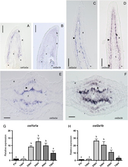
Collagens type II alpha 1 b and type I alpha 1 are constituents of the fin skeleton. (A–F) In situ hybridization of col1a1 (A, C and E) and col2a1b (B, D and F) in longitudinal (A–D) and transversal (E–F) sections of regenerated fins. A–B: Ray blastema at 2.5 dpa. C–D: Ray blastema at 3 dpa. E–F: Ray blastema at 7 dpa. Bars represent 25 μm (E–F) and 50 μm (A–D). Arrowhead is actinotrichia. Asterisk is lepidotrichia. e is epidermis. b is basal epidermal layer. c is connective tissue. (G–H) Expression analysis of col1a1a and col2a1b. Total RNA was extracted from different stages of fin regeneration and the relative transcript levels of col1a1a and col2a1b were determined by qRT-PCR. col1a1a (G), col2a1b (H) in different regeneration stages: Fin, non regenerating fin; 1 dpa, 1 days post amputation; 2 dpa, 2 days post amputation; 3 dpa, 3 days post amputation; 4 dpa, 4 days post amputation; 7 dpa, 7 days post amputation. Bars represent the mean of two independent biological samples ± SE. Different letters indicate a significant difference between samples according to the corresponding ANOVA (P < 0.05). |
| Genes: | |
|---|---|
| Fish: | |
| Condition: | |
| Anatomical Term: | |
| Stage: | Adult |
Reprinted from Developmental Biology, 354(1), Durán, I., Marí-Beffa, M., Santamaría, J.A., Becerra, J., and Santos-Ruiz, L., Actinotrichia collagens and their role in fin formation, 160-172, Copyright (2011) with permission from Elsevier. Full text @ Dev. Biol.