
Fig. 3
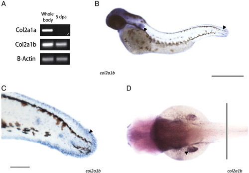
col2a1b expression. (A) Semiquantitative RT-PCR of col2a1a and col2a1b in the whole body of 1 month postfertilization (left) and regenerating fin (rigth) tissues. b-actin is used as a control of constitutive expression. (B–D) In situ hybridization of a zebrafish embryo at 48 h stage. B. Whole mount in situ hybridization of col2a1b gene. Arrows point at the pectoral and tail fin folds. C. Detail of col2a1b gene expression at the tail bud. Note the expression at neuromast cells along the prospective lateral line. Arrow points at the distal portion of the tail fin fold. D. Dorsal view of col2a1b gene expression. Note the expression at the forebrain and midbrain. Arrow points to a labeled left pectoral fin bud. Bars represent 0.5 mm (B and D) and 0.2 mm (B). |
| Genes: | |
|---|---|
| Fish: | |
| Condition: | |
| Anatomical Terms: | |
| Stage Range: | Long-pec to Days 30-44 |
Reprinted from Developmental Biology, 354(1), Durán, I., Marí-Beffa, M., Santamaría, J.A., Becerra, J., and Santos-Ruiz, L., Actinotrichia collagens and their role in fin formation, 160-172, Copyright (2011) with permission from Elsevier. Full text @ Dev. Biol.