|
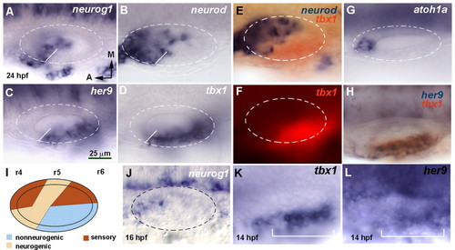
her9 and tbx1 expression is complementary to the neurogenic domain. (A-H,J-L) Dorsal views of flat-mounted zebrafish embryos at 24 hpf, with anterior to the left and medial to the top. Dashed circles delineate the otic vesicle. In situ hybridization was for neurog1 (A), neurod (B), her9 (C), tbx1 (D) and atoh1a (G). White lines in A-D indicate the boundary between the neurogenic and non-neurogenic domains. Double in situ hybridization is shown for tbx1 (red chromogen in E and H, red fluorescence in F) and neurod (blue, E) or her9 (blue, H). (J) neurog1 expression at 16 hpf. (K,L) Expression of tbx1 and her9 in the otic placode at 14 hpf. The bracket indicates the extent of the placodal domain. (I) Schematic representation of the neurogenic, sensory and non-neurogenic territories. r, rhombomere. All images are at the same magnification.
|