
Fig. 7
- ID
- ZDB-FIG-110121-35
- Publication
- Yu et al., 2011 - The Cell Adhesion-associated Protein Git2 Regulates Morphogenetic Movements during Zebrafish Embryonic Development
- Other Figures
- All Figure Page
- Back to All Figure Page
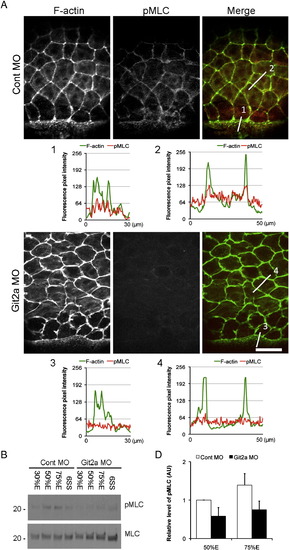
Git2 functions through myosin II-dependent contractility to regulate epiboly. (A) Immunofluoresence staining of phosphorylated Myosin light chain (pMLC) and F-actin in EVL cells of control and git2a morphants at 75% epiboly. In control embryos, pMLC (red) co-localized with cortical actin (green) at the cell periphery in EVL cells and at the margin where the EVL contacts the YSL. However, a significant reduction of pMLC staining was observed in git2a morphants. Scale bar, 50 μm. The F-actin and pMLC fluorescence pixel intensity profiles of EVL cells in control (1, 2) and git2a morphant (3, 4) embryos was aligned. (B) Western blotting of pMLC and total levels of MLC in control and git2a morphants from 30% epiboly to the 6 somite stage. (C) Quantification of the relative level of pMLC: total MLC from control and git2a morphants at 50% and 75% epiboly from three independent experiments. An arbitrary unit (AU) is designated as the pMLC level at 50% epiboly. |
| Antibody: | |
|---|---|
| Fish: | |
| Knockdown Reagent: | |
| Anatomical Terms: | |
| Stage Range: | 50%-epiboly to 5-9 somites |
Reprinted from Developmental Biology, 349(2), Yu, J.A., Foley, F.C., Amack, J.D., and Turner, C.E., The Cell Adhesion-associated Protein Git2 Regulates Morphogenetic Movements during Zebrafish Embryonic Development, 225-237, Copyright (2011) with permission from Elsevier. Full text @ Dev. Biol.