
Fig. S2
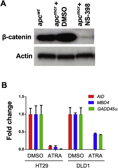
Knockdown of β-Catenin Levels by Cox-2 Inhibitor and Rescue of Demethylase by Retinoic Acid, Related to Figure 2 (A) Western blot showing levels of β-catenin in apcwt and apcmcr zebrafish embryos (72hpf) treated with DMSO (vehicle) or NS-398 (Cox-2 inhibitor, 10 μM). Actin was used as a loading control. (B) RT-PCR showing expression of AID, GADD45α and MBD4 in HT29 and DLD1 cells treated with vehicle (DMSO) or ATRA (1μM). y axis values are fold changes in expression of indicated genes in ATRA treated cells normalized first to 28S levels and then to mRNA/28S ratio from DMSO treated cells, valued as 1. Error bars are +/- SD. |
Reprinted from Cell, 142(6), Rai, K., Sarkar, S., Broadbent, T.J., Voas, M., Grossmann, K.F., Nadauld, L.D., Dehghanizadeh, S., Hagos, F.T., Li, Y., Toth, R.K., Chidester, S., Bahr, T.M., Johnson, W.E., Sklow, B., Burt, R., Cairns, B.R., and Jones, D.A., DNA demethylase activity maintains intestinal cells in an undifferentiated state following loss of APC, 930-942, Copyright (2010) with permission from Elsevier. Full text @ Cell