
Fig. S1
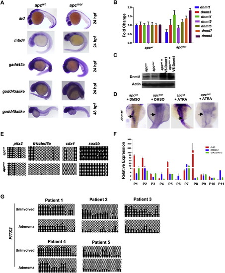
Expression of Demethylase Genes and dnmt1 and Verification of Hypomethylated Genes in apc Mutant Zebrafish Embryos, Related to Figure 1 (A) Whole mount in situ staining for aid, mbd4, gadd45α and gadd45αlike in apc mutants (apcmcr) and siblings (apcwt) embryos. Lateral view is shown for 24hpf and 48hpf old embryos. (B) RT-PCR measuring levels of all zebrafish DNA methyltransferases in apcmcr and apcwt embryos. y axis shows fold change normalized to 28S levels first and then to dnmt/28 s ratio from apcwt, valued as 1. (C) Western blots showing Dnmt1 levels in apcmcr and apcwt embryos injected with V5-Dnmt1 plasmid. Upper panel is probed with an antibody generated against zebrafish Dnmt1. Actin is used as a loading control. (D) Whole mount in situ staining for dnmt1 in apcmcr and apcwt embryos treated with DMSO or ATRA (1μM). Black arrow points to the expression in intestine. (E) Circle diagram showing bisulfite sequencing results for pitx2, frizzled8, cdx4 and sox9b promoter in apcmcr and apcwt embryos at 72hpf. (F) RT-PCR showing expression of AID, GADD45α and MBD4 in adenoma tissues isolated from FAP patients (patients bearing mutations in APC gene). P1 through P11 refers to patient′s sample ID. y axis values are fold changes in expression of indicated genes in adenoma tissues normalized first to 28S levels and then to mRNA/28S ratio from matching uninvolved tissue, valued as 1. Error bars are +/- SD. (G) Circle diagram showing bisulfite sequencing results for PITX2 promoter in adenoma and normal-appearing tissue from five different FAP patient samples. Circles represent methylation status of cytosines which are located in CpG contexts. Unmethylated cytosines are shown by open circle whereas closed circles depict methylated cytosines. |
Reprinted from Cell, 142(6), Rai, K., Sarkar, S., Broadbent, T.J., Voas, M., Grossmann, K.F., Nadauld, L.D., Dehghanizadeh, S., Hagos, F.T., Li, Y., Toth, R.K., Chidester, S., Bahr, T.M., Johnson, W.E., Sklow, B., Burt, R., Cairns, B.R., and Jones, D.A., DNA demethylase activity maintains intestinal cells in an undifferentiated state following loss of APC, 930-942, Copyright (2010) with permission from Elsevier. Full text @ Cell