
Fig. 12
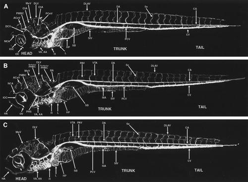
Circulation in the developing zebrafish at 5.5–7.5 days postfertilization (dpf). (A) Angiogram of a developing zebrafish at approximately 5.5 dpf, compiled from five separate reconstructions pasted together. (B) Angiogram of a developing zebrafish at approximately 6.5 dpf, compiled from five separate reconstructions pasted together. (C) Angiogram of a developing zebrafish at approximately 7.5 dpf, compiled from six separate reconstructions pasted together. All panels are oriented with rostral to the left and ventral down, and particular vessels have been labeled in each panel. A glossary of the names corresponding to all labeled vessels is provided in Table 1. |
Reprinted from Developmental Biology, 230(2), Isogai, S., Horiguchi, M., and Weinstein, B.M., The vascular anatomy of the developing zebrafish: an atlas of embryonic and early larval development, 278-301, Copyright (2001) with permission from Elsevier. Full text @ Dev. Biol.