
Fig. 2
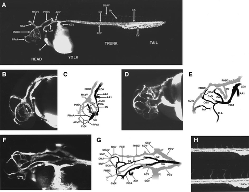
Circulation in the developing zebrafish at 1.2–1.5 days postfertilization (dpf). (A) Angiogram of a developing zebrafish at approximately 1.5 dpf, compiled from two separate reconstructions pasted together. Lateral view. (B) Angiogram of the head of a developing zebrafish at approximately 1.3 dpf. Lateral view. (C) Diagram of vessels in (B). Only the vessels on the left side are shown. (D) Angiogram of the head of a developing zebrafish at approximately 1.5 dpf. Ventral–rostral–lateral view. (E) Diagram of vessels in (D). Only the vessels on the left side are shown. (F) Angiogram of the head and cranial trunk of a developing zebrafish at approximately 1.5 dpf. Dorsal–lateral view. (G) Diagram of vessels in (F). (H) Angiogram of the caudal trunks of developing zebrafish at approximately 1.2–1.5 dpf. Lateral views. The emergence of the intersegmental (intersomitic) vessels of the trunk is shown. The upper angiogram shows a somewhat earlier stage than the lower angiogram. All lateral views are from the left side. A glossary of the names corresponding to all labeled vessels is provided in Table 1. |
Reprinted from Developmental Biology, 230(2), Isogai, S., Horiguchi, M., and Weinstein, B.M., The vascular anatomy of the developing zebrafish: an atlas of embryonic and early larval development, 278-301, Copyright (2001) with permission from Elsevier. Full text @ Dev. Biol.