
Fig. 4
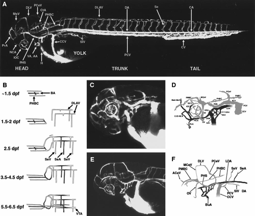
Circulation in the developing zebrafish at approximately 2.5 days postfertilization (dpf). (A) Angiogram of a developing zebrafish at approximately 2.5 dpf, compiled from three separate reconstructions pasted together. Lateral view, labeled. (B) Schematic diagram showing vessels of the caudal head and cranial trunk, how these vessels become connected, and how this changes over the course of several days. (C) Angiogram of the head of a developing zebrafish at approximately 2.5 dpf. Ventral–lateral view. (D) Diagram of vessels in (C). Only the vessels on the left side are shown, for clarity. AA1–AA4 have formed, and the PHS is now on line. (E) Angiogram of the head of a developing zebrafish at approximately 2.5 dpf. Lateral view. (F) Diagram of vessels in (E). Only the vessels on the left side are shown, for clarity. All six aortic arches are now carrying blood flow. All panels are oriented with rostral to the left, and all lateral views are from the left side. A glossary of the names corresponding to all labeled vessels is provided in Table 1. |
Reprinted from Developmental Biology, 230(2), Isogai, S., Horiguchi, M., and Weinstein, B.M., The vascular anatomy of the developing zebrafish: an atlas of embryonic and early larval development, 278-301, Copyright (2001) with permission from Elsevier. Full text @ Dev. Biol.