
Fig. 8
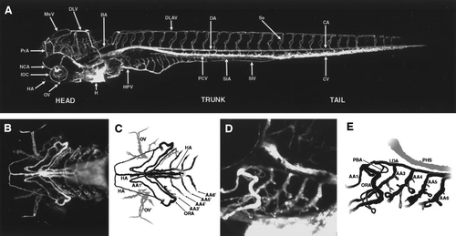
Circulation in the developing zebrafish at approximately 4 days postfertilization (dpf). (A) Angiogram of a developing zebrafish at approximately 4 dpf, compiled from five separate reconstructions pasted together. Lateral view, labeled. (B) Angiogram of the ventral head of a developing zebrafish at approximately 4 dpf. (C) Diagram of vessels in (B). In this ventral view only the aortic arches and adjacent vessels are visible. All extant aortic arches are shown, as well as the hypobranchial vessels. (D) Higher magnification (20x) angiogram of the ventral head of a developing zebrafish at approximately 4 dpf. Lateral view, showing primarily aortic arches. (E) Diagram of vessels in (D). Note the vascular loops associated with AA3–AA6. Separation of these AA into parallel afferent and efferent derivatives (ABA and ABF) is in progress but not well visualized in this particular orientation of the embryo. In all panels rostral is to the left, and all lateral views are from the left side. A glossary of the names corresponding to all labeled vessels is provided in Table 1. |
Reprinted from Developmental Biology, 230(2), Isogai, S., Horiguchi, M., and Weinstein, B.M., The vascular anatomy of the developing zebrafish: an atlas of embryonic and early larval development, 278-301, Copyright (2001) with permission from Elsevier. Full text @ Dev. Biol.