
Fig. 3
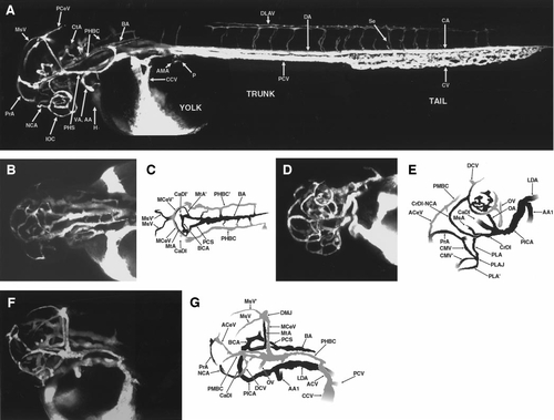
Circulation in the developing zebrafish at approximately 2 days postfertilization (dpf). (A) Angiogram of a developing zebrafish at approximately 2 dpf, compiled from three separate reconstructions pasted together. Lateral view, labeled. (B) Angiogram of the head of a developing zebrafish at approximately 1.8 dpf. Dorsal view. (C) Diagram of vessels in (B). Vascular connections are apparent between the BA and both PHBC. (D) Angiogram of the head of a developing zebrafish at approximately 2.0 dpf. Ventral–rostral–lateral view. (E) Diagram of vessels in (D). Only the vessels on the left side are shown, for clarity. Note that the PPrA has now disconnected from the CrDI and the PMsA has disconnected from the PMBC. (F) Angiogram of the head of a developing zebrafish at approximately 2.0 dpf. Dorsal–lateral view. The vessels shown are mostly those on the left side, for clarity. The major arterial and venous pathways of the head at this stage are apparent. (G) Diagram of vessels in (F). All panels are oriented with rostral to the left, and all lateral views are from the left side. A glossary of the names corresponding to all labeled vessels is provided in Table 1. |
Reprinted from Developmental Biology, 230(2), Isogai, S., Horiguchi, M., and Weinstein, B.M., The vascular anatomy of the developing zebrafish: an atlas of embryonic and early larval development, 278-301, Copyright (2001) with permission from Elsevier. Full text @ Dev. Biol.