
FIGURE
Fig. 1
Fig. 1
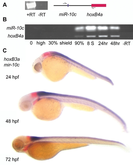
Spatial and temporal expression profile of miR-10c. A) RT-PCR with primers located 5′ of miR-10c and within the coding region of exon 1 of HoxB4a shows inclusion of miR-10c and HoxB4a on the same transcript. PCR 35 cycles, -RT: no reverse transcriptase added. B) RT-PCR shows similar temporal expression during development of HoxB4a (28 cycles) and miR-10c pre-miRNA (35 cycles). C) Whole mount in situ hybridization on different stage Zebrafish embryos shows mutually exclusive expression of the HoxB3a rhombomere 5/6 domain (red) with miR-10c (purple). |
Expression Data
| Genes: | |
|---|---|
| Fish: | |
| Anatomical Terms: | |
| Stage Range: | 90%-epiboly to Protruding-mouth |
Expression Detail
Antibody Labeling
Phenotype Data
Phenotype Detail
Acknowledgments
This image is the copyrighted work of the attributed author or publisher, and
ZFIN has permission only to display this image to its users.
Additional permissions should be obtained from the applicable author or publisher of the image.
Full text @ PLoS One