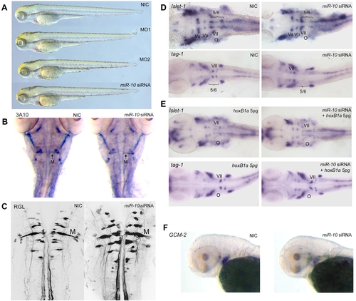
Overexpression of miR-10 induces HoxB1a and HoxB3a loss of function phenotypes. A) Wildtype (WT), miR-10 morphant (MO1, MO2) and miR-10 siRNA overexpression embryos at 72 hpf show no apparent developmental differences. B) Mauthner neuron development as visualized by 3A10 neurofilament immunostaining in 72 hpf embryos shows no differences between miR-10 siRNA injected embryos and controls. C) Confocal images of reticulospinal hindbrain neurons in retrograde labeled, 5 day old embryos. Wildtype and miR-10 siRNA injected embryos are similar. D) Islet-1 and tag-1 in situ hybridization on 30 hpf wildtype and miR-10 siRNA injected embryos. Flatmounts of head regions are shown. In wildtype embryos the VIIth cranial nerve migrates into rhombomere 5/6 at the level of the otic vesicle. In miR-10 siRNA injected embryos the VIIth nerve does no longer migrate out of rhombomere 4. E) Co-injection of 5pg HoxB1a RNA rescues the miR-10 siRNA induced migration defect of the VIIth cranial nerve as shown by islet-1 and tag-1 in situ hybridization. F) Gcm-2 expression is downregulated in miR-10 siRNA injected embryos, which is consistent with repression of HoxB3a.
|
