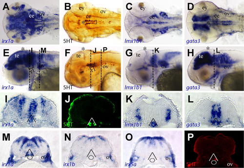
Expression of irx1a in hindbrain serotonergic neurons. A-H: Dorsal view of flat-mounted (A-D) and a lateral view (E-H) of a 48 hpf zebrafish brain region. A,B,E,F: irx1a is expressed in two rows of cells in rostral ventral hindbrain (black brackets, A,E) and have the same pattern as serotonergic neurons (B,F). C,D,G,H: The serotonergic markers Lmx1b1 (C,G) and Gata3 (D,H) are also expressed in the rostral ventral hindbrain. I-L: Transverse section of zebrafish hindbrain at rhombomere 1 position which are indicated in E-H, respectively. J: The serotonergic neurons are located in the ventral hindbrain (dashed lines) and irx1a is expressed in the same region. K: Lmx1b1 is expressed in the serotonergic neurons as well as in the floor plate. On the other hand, Gata3 is expressed in the serotonergic neurons but not the floorplate, and note that the ventral expression domain of Gata3 is boarder than the serotonergic neurons. M,P: Transverse section of zebrafish hindbrain at the otic vesicle region, which is indicated in E,F, respectively. The ventral expression domain of irx1a is on the boundary of notochord (dashed line circles in M) and is overlapped with serotonergic neurons (P). N,O: The other iro1 homologue irx1b (N) nor irx5a (O) are not expressed in this domain (N).
|
