
Fig. 2
- ID
- ZDB-FIG-070802-4
- Publication
- Tessmar-Raible et al., 2007 - Conserved sensory-neurosecretory cell types in annelid and fish forebrain: insights into hypothalamus evolution
- Other Figures
- All Figure Page
- Back to All Figure Page
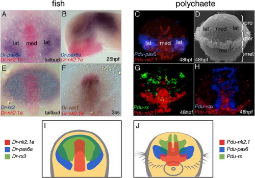
Conserved Regionalization of the Zebrafish and Platynereis Forebrain (A, E, and F) Dorsal and (B) lateral views of zebrafish embryos hybridized with the indicated riboprobes; stages as indicated. (A, E, and F) Anterior to the top and (B) to the left, (B) eyes removed. (C, G, and H) Apical views of Platynereis embryos hybridized with the indicated riboprobes. (D) SEM picture of a Platynereis larva. Pro, prostomium; per, peristomium; met, metastomium; mo, mouth. (I and J) Expression synopsis of zebrafish nk2.1a, pax6.1, and rx3 (I) and of the polychaete orthologs (J). Yellow, anterior neuroectoderm; med, medial; lat, lateral forebrain. |
| Genes: | |
|---|---|
| Fish: | |
| Anatomical Terms: | |
| Stage Range: | 1-4 somites to Prim-5 |
Reprinted from Cell, 129(7), Tessmar-Raible, K., Raible, F., Christodoulou, F., Guy, K., Rembold, M., Hausen, H., and Arendt, D., Conserved sensory-neurosecretory cell types in annelid and fish forebrain: insights into hypothalamus evolution, 1389-1400, Copyright (2007) with permission from Elsevier. Full text @ Cell