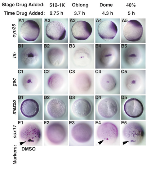
Nodal signals pattern the dorsal mesoderm and endoderm along the animal-vegetal axis in a time-dependent manner. Dorsal cell fates were examined in embryos treated with DMSO (A1-E1), or with SB-431542 at various time points. (A2-5) cyp26 expression was expressed at the margin in embryos treated at MBT, but is expressed in more animal locations at later time points. (B2-5) flh is first detected in embryos treated at 3.7 h. (C2-5) gsc is first observed in embryos treated at 4.3 h (dome stage), but is expressed at normal levels in embryos treated after 5 h (40% epiboly). mezzo transcripts are observed in embryos treated after 5 h (40% epiboly) (D5), but not at earlier stages (D2-4). sox17 is expressed in the dorsal forerunner cells in embryos treated 4.3 h (dome stage) (E4, arrowhead), but is first detected in endoderm progenitors in embryos treated at 5 h (40% epiboly) (E5). Lateral views of embryos at 10 h are depicted in A1-5, dorsal to the right. Dorsal views of embryos at 7 h (60% epiboly) (B1-C5), 5.5 h (germ ring) (D1-5) and 8 h (80% epiboly) (E1-5) are depicted. Arrowheads (E1, 4, 5) indicate sox17 in dorsal forerunner cells. All embryos are siblings.
|
