|
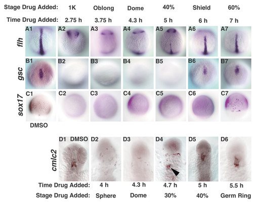
Cell fate specification is delayed squint mutants. Cell fates were examined in sqt mutant embryos treated with DMSO (A1-D1), or with SB-431542 at various time points. (A1-7) flh was first expressed at the midline in embryos treated at 5 h (A5). (B2-7) gsc expression is first detected in embryos treated at 6 h (B6). (C2-7) sox17 expression is first detected when embryos are treated at 7 h (C7). (D1-7) cmlc2 expression was first detected in embryos treated 4.7 h (D4, arrowhead). Dorsal views of 10 h (A1-B7), 8 h (C1-C7) or 24 h (D1-D6). In D1-D6, anterior is up. The embryos in Figs. 8 and 9 are from the same clutch and weretreated in parallel, along with wild type controls (not shown).
|