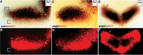
Localization of eng3 and wnt1 (blue) relative to pax2.1 (red/fluorescent) in double in situ hybridizations. All embryos are oriented anterior to the top; insets give the area shown in detail, and arrowheads point to identical landmark cells in corresponding panels. (A) 90% epiboly stage embryo stained for wnt1/pax2.1. The two brackets mark the area where only pax2.1 or wnt1 are expressed. The wnt1 domain extends further laterally than pax2.1, whereas pax2.1 extends further posteriorly; at this stage the anterior boundaries of wnt1 and pax2.1 coincide. (B) Fluorescence image of A. FastRed fluorescence is quenched in the overlapping part by the wnt1 signal, but not posteriorly. (C) 90% epiboly stage embryo stained for eng2/pax2.1. The eng2 domain lies within the pax2.1 domain. (D) The fluorescent image of C, clearly showing that the eng2 cells lie within the pax2.1 domain. (E,F) 1-somite embryo stained for eng3/pax2.1; the initial eng3 expression domain lies within the pax2.1 domain; shortly after, the two domains become coincident (not shown).
|
