|
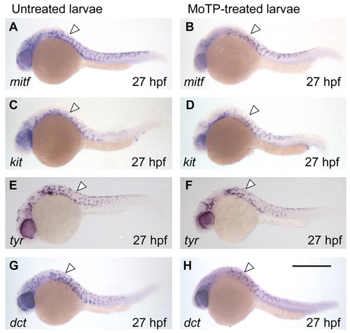
Melanocyte lineages develop to late stages in the presence of MoTP. (A-H) The development of melanocyte lineages in MoTP-treated (B,D,F,H) and untreated (A,C,E,G) larvae were examined by whole-mount RNA in situ hybridization with mitf (A,B), kit (C,D), tyr (E,F) and dct (G,H). For these experiments, embryos were incubated in MoTP solution from 14 to 27 hpf, then immediately fixed for in situ analysis. The developmental patterns and the numbers of cells (white arrowheads) for each of the markers are indistinguishable in the MoTP-treated larvae from those in untreated larvae. Untreated larvae were reared in PTU to completely block melanin synthesis, thereby allowing easier visualization of NBT/BCIP precipitates in melanocytes. Scale bar: 500 μm.
|