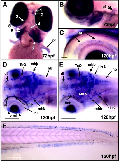
Whole-mount in situ hybridization: 72-120 hours postfertilization (hpf). A: Ventral view, 72 hpf. Although Cx43α transcripts are difficult to detect in the ear and heart by 72 hpf, they are readily detected in all seven pharyngeal arches (numbered) and in the pectoral fin cartilage (pf). B: Lateral view of the same embryo as in A (pf, pectoral fin). C: Lateral view, 120 hpf. Expression can be seen in the inner nuclear layer (inl) of the retina. D: Oblique dorsal view, 120 hpf. Cx43α expression can be identified in the diencephalon (di), optic tectum (TeO), the hindbrain (hb), and in the boundaries between these regions of the brain, i.e., the forebrain-midbrain boundary (fmb) and the midbrain-hindbrain boundary (mhb). inl, inner nuclear layer; v, ventral retina. E: Oblique dorsal view, 120 hpf. Plane of focus is more dorsal than in D. Cx43α also delineates the rhombomere 1-rhombomere 2 boundary (r1-r2); the region between the midbrain-hindbrain boundary (mhb) and the r1-r2 is the cerebellum. Expression in the fourth ventricle (4th v) persists. Other abbreviations as in D. F: Lateral view, 120 hpf. Cx43α expression in the median fin fold cartilage. Scale bars = 120 μm in A, 125 μm in B, 35 μm in C, 250 μm in D,E, 140 μm in F.
|
