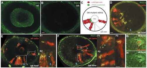
RGC neurogenesis in the absence of RGC-derived signals. (A,B) Zn5 staining of wild-type (A) and lak mutants (B) at 55 hpf. Mutant retina is devoid of RGCs. (C) Schematic showing predicted results of transplant experiments, based on the sequential-induction model. Red cells represent wild-type clones in a lak mutant retina. The clone marked 1 is predicted to give rise to RGCs (green) because it is near the optic stalk (dashed circle). Clone 2 is predicted to give rise to RGCs because it is near clone 1, which contains earlier-born RGCs. Clone 3 is located far from the optic stalk and from RGCs; the model predicts it should fail to give rise to RGCs. (D-F) Wild-type-into-lak chimeras. (D) Only wild-type cells (red) give rise to zn5+ RGCs (green). (E) A wild-type-into-lak chimera that challenges the sequential-induction model. One clone (E1) is completely surrounded by mutant tissue and yet still forms RGCs (green). (E1, E2) High-magnification views of indicated boxed areas, showing expression of RGC markers (green) by wild-type cells (arrowhead). Both are at the same scale. (F) A wild-type-into-lak chimera in which donor cells are present only in temporal retina. These clones give rise to zn5+ RGCs. (F1-F3) High-magnification views of boxed areas (all are at the same scale). In F1, an axon (arrowheads) extends from the double-labeled RGC cell body (arrow). (G,H) A lak-into-wild-type chimera, showing that ath5 is cell-autonomously required for RGC formation. Mutant donor cells (red; arrowhead) are the only cells in the GCL not labeled with zn5 (green). (H) The same field of view, showing zn5 alone to illustrate the gap in zn5 expression where the lak cells are (arrowhead). Anterior/nasal is left and dorsal up in all images. The blue arrow indicates the choroid fissure. S, donor-derived skin cells, not in neural retina; wt, wild type. Scale bars: in B,D, 20 µm for A,B,D,E,F; 5 µm for E2,F3,H.
|
