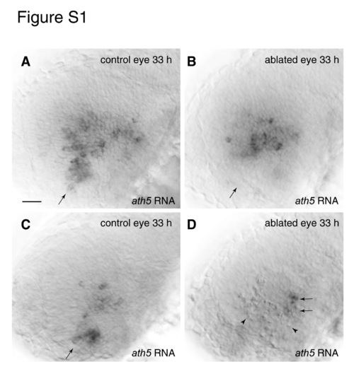
Laser ablation of ventronasal retina at 22-28 somites fails to block the ath5 wave. Laser ablation of the ventronasal retinal quadrant was performed on Pax6-DF4:mGFP larvae ranging in age from 22 to 28 somites. The larvae were subsequently sacrificed at 30 hpf (not shown) or 33 hpf (A-D) and stained for ath5 mRNA. All 30 hpf animals lacked ath5 expression in the ablated (but not the intact) eye, indicating that onset of ath5 expression was blocked efficiently in this experiment (n=6; not shown). (A,B) Control (A) and laser-ablated (B) eyes from the same 33 hpf larva. This individual is a representative sample from this experiment – all larvae showed essentially the same phenotype (n=12). In control retinae (A), ath5 is expressed in the ventronasal patch (arrow) and in central retina. In ablated retinae (B), ath5 expression is eliminated from ventronasal retina (arrow) but is expressed normally by central retina cells, suggesting that early ablation of the ventronasal patch does not prevent central retinal cells from expressing ath5 on time. Pyknotic cells were observed in ventronasal retina but are out of the plane of focus of the image in B. (C,D) Control (C) and ablated (D) eyes from another individual in the same experiment. In this individual the ablated region encompasses a much larger area of the retina than was intended. In addition to ventronasal retina, much of the anterior region of central retina has been killed, as indicated by the high density of pyknotic nuclei (D; arrowheads). Nevertheless, ath5 is expressed normally by cells in the temporal half of central retina (arrows). The ath5-expressing cells are just adjacent to the temporal-most extent of the laser-targeted region, as shown by the close apposition of pyknotic and ath5-expressing cells. Thus, joint ablation of ventronasal and antero-central retina does not prevent the ath5 wave from initiating in the temporal half of the retina. Anterior is leftwards and dorsal upwars in all panels. Scale bar: 20 μm.
|
