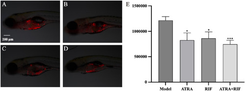
Growth of zebrafish tumor cells (HL - 60) after sample treatment (A) Model control group; (B) ATRA treatment group; (C) RIF treatment group; (D) ATRA + RIF combined treatment group. Compared with the model control group, the fluorescence intensity of tumor cells in the ATRA group and the RIF group decreased significantly (*p < 0.05), and that in the ATRA + RIF group decreased significantly (***p < 0.001). (E) Statistical analysis chart of the fluorescence intensity of tumor cells (pixels), and the data are expressed as mean ± SE.

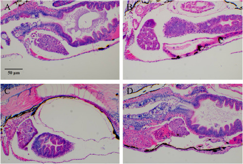
Typical images of zebrafish tumor cell (HL-60) migration after RIF intervention. (A) Model group; (B–D) ATRA, RIF, and ATRA + RIF. In the liver tissue of zebrafish from the model control group, extensive fat vacuolar degeneration (indicated by yellow arrows) is observed, confirming the successful establishment of the model. In contrast, the liver tissue of zebrafish in the treatment groups shows a significant reduction in fat vacuolar degeneration, suggesting that ATRA and Compound Huangdai Tablets have a potential protective effect against liver injury.

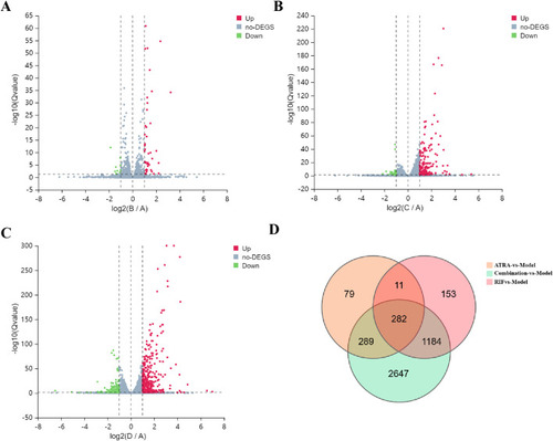
Transcriptomic profiling of differentially expressed genes (DEGs) across treatment groups. (A–C) Volcano plots show DEGs under |log2FC| ≥ 1 and Q-value ≤0.05 thresholds for (A) ATRA-vs-Model (45 upregulated, red; 10 downregulated, green), (B) RIF-vs-Model (168 upregulated, red; 29 downregulated, green), and (C) Combination-vs-Model (385 upregulated, red; 189 downregulated, green). Dashed lines demarcate significance thresholds. (D) Venn diagram illustrates overlapping DEGs (n = 282) common to all three comparisons, indicating shared transcriptional perturbations.

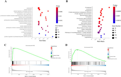
Integrated mechanisms of ATRA in APL therapy. Functional analyses identify key pathways mediating ATRA’s therapeutic effects: (A) GO analysis links ATRA to muscle contraction, creatine synthesis, and coagulation; (B) KEGG analysis highlights altered FoxO and PI3K-Akt signaling alongside apoptotic regulation; Gene Set Enrichment Analysis (GSEA) further validates FoxO (C) and apoptosis (D) as critical regulatory axes.

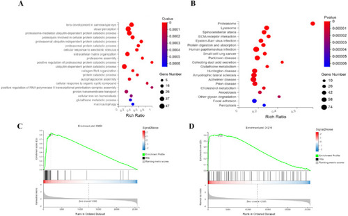
Synergistic targeting of proteasome and ferroptosis pathways underlies RIF’s anti-leukemic efficacy. Functional dissection identifies RIF-driven pathway perturbations: (A) GO analysis reveals coordinated dysregulation in ubiquitin-proteasome system (proteasome assembly, ubiquitin-dependent/independent degradation) coupled with autophagic flux and iron homeostasis; (B) KEGG analysis confirms proteasome activation (intracellular proteostasis hub) and ferroptosis-glutathione axis alterations; Gene Set Enrichment Analysis (GSEA) validates coherent modulation of (C) Proteasome and (D) Ferroptosis pathways. These findings highlight a dual-pathway mechanism where RIF suppresses leukemia progression by inhibiting proteasome hyperactivity and inducing iron-dependent redox stress.

Mechanism of the combination therapy in synergistically suppressing APL through triple-axis targeting of the proteasome, ferroptosis, and lysosome pathways. (A) GO analysis reveals marked perturbations in ubiquitin-proteasome system (UPS) processes, including ubiquitin-dependent/independent proteolysis, proteasome assembly, and enhanced proteasomal degradation regulation, alongside disrupted iron ion homeostasis and autophagosome dynamics. (B) KEGG pathway enrichment indicates significant activation of the proteasome pathway (central to proteostasis), ferroptosis, glutathione metabolism (linked to oxidative stress), and concurrent enrichment of neurodegenerative disease pathways (e.g., Parkinson’s and Alzheimer’s). (C,D) GSEA validates robust modulation of the proteasome and lysosome pathways.

Acknowledgments
This image is the copyrighted work of the attributed author or publisher, and ZFIN has permission only to display this image to its users. Additional permissions should be obtained from the applicable author or publisher of the image. Full text @ Front Pharmacol