
- Title
-
Piezo 1 and Piezo 2 in the Chemosensory Organs of Zebrafish (Danio rerio)
- Authors
- Aragona, M., Mhalhel, K., Cometa, M., Franco, G.A., Montalbano, G., Guerrera, M.C., Levanti, M., Laurà, R., Abbate, F., Vega, J.A., Germanà, A.
- Source
- Full text @ Int. J. Mol. Sci.
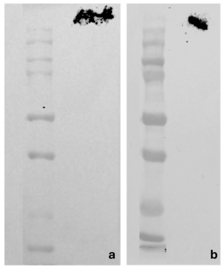
Western blot analyses showing bands corresponding to the molecular weights of the zebrafish Piezo 1 (a) and Piezo 2 (b) proteins. |
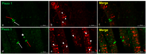
(a–f) Zebrafish olfactory lamellae, dorsal view. We conducted an immunohistochemical detection (using the immunofluorescences method) of Piezo 1 in a colocalization view with Calretinin as a specific marker for the neuronal subpopulation cells. (a,d) The kappe cells (red arrows) and microvillous sensory cells (white arrows) were immunopositive to Piezo 1. (d) Piezo 1 was localized in crypt neurons (gallon arrow). (b,e) The ciliated sensory cells were immunolabeled to Calretinin (arrowhead). (c,f) A colocalization view that shows no overlap in the labelling. Magnification, 40×; scale bar, 20 µm. EXPRESSION / LABELING:
|
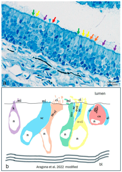
The olfactory epithelium of an adult zebrafish. (a) Photomicrographs of the semithin section showing the kappe cells (purple arrows), one-ciliated ON (yellow arrows), multi-ciliated ON (orange arrows), microvillous ON (green arrows), and crypt neurons (red arrow) surrounded by special supporting cells (blue arrowheads). Toluidine blue; magnification, 20×. (b) A zebrafish epithelium olfactory neuron’s graphic representation modified using the transmission electron microscopy micrograph from our previous study [10]. Abbreviations: k, kappe cells; sc, supporting cells; m-ci, multi-ciliated ON; n, nucleus; o-ci, one-ciliated ON, ci, cilium; mv, microvillous ON; mi, microvillus; cn, crypt neurons, with several cilia within the crypt (asterisk), special supporting cells (sc), and basal lamina (bl) |
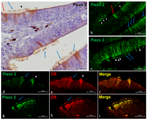
Piezo 2 immunolocalizations in zebrafish olfactory lamellae, dorsal view. (a) The immunohistochemical detection (using a peroxidase method, Haematoxylin-stained) of Piezo 2 showing kappe neurons (red arrow), one-ciliated sensory cells (arrowhead), and multi-ciliated sensory cells (blue arrows). (b,c) The Piezo 2 immunolocalization showing kappe neurons (red arrow), one-ciliated sensory cells (arrowhead), and multi-ciliated sensory cells (blue arrows). (d,e,g,h) The immunohistochemical detection of Piezo 2 and Calretinin showing one-ciliated (arrowhead) and multi-ciliated (arrows) sensory cells. (f,i) A Piezo 2–Calretinin colocalization view. Magnification, 40×; scale bar, 20 µm EXPRESSION / LABELING:
|
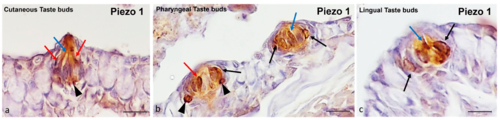
Zebrafish taste buds, transversal view. The immunohistochemical detection (using a peroxidase method) of Piezo 1. (a) In the cutaneous taste buds, the Merkel-like cells (arrowheads), light cells (blue arrows), and dark cells (red arrows) showed immunopositivity to Piezo 1. (b) In the pharyngeal taste buds, the Merkel-like cells (arrowheads), light cells (blue arrows), dark cells (red arrows), and supporting cells (black arrows) showed immunoreactivity to Piezo 1. (c) In the lingual taste buds, the light cells (blue arrow) and supporting cells (black arrows) were Piezo 1-immunolabelled. Magnification, 40×; scale bar, 20 µm. EXPRESSION / LABELING:
|
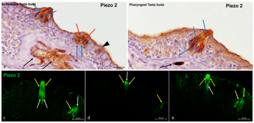
Zebrafish taste buds, transversal view. (a,b) The immunohistochemical detection (using a peroxidase method, Haematoxylin-stained) of Piezo 2. The light cells (blue arrows), dark cells (red arrows), and nerve cells (black arrows) were Piezo 2-immunopositive. (a) Isolated chemosensory cells (black arrowhead) were immunoreactive to Piezo 2. (c–e) The multiple focal planes of the Piezo 2 immunohistochemical detections (using a fluorescence method). The light cells (white arrows) and dark cells (yellow arrows) were immunoreactive to Piezo 2. Magnification, 40×; scale bar, 20 µm. EXPRESSION / LABELING:
|
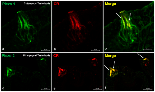
Zebrafish taste buds, transversal view. The immunohistochemical detection (using fluorescence methods) of Piezo 1 and Piezo 2. (a–c) The double experiments with Piezo 1 and Calretinin showed an overlapping stain in the sensory cells (arrows) of the cutaneous taste buds. (d–f) The double experiments for Piezo 2 and Calretinin showed an overlapping stain in the sensory cells (arrows) of the pharyngeal taste buds. Magnification, 40×; scale bar, 20 µm. EXPRESSION / LABELING:
|
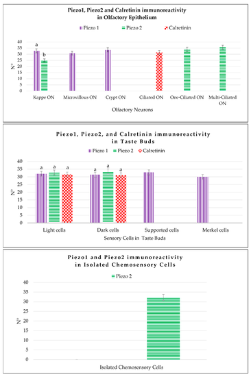
Graphical representation of the immunoreactive cell counts: the kappe olfactory neurons (Kappe ON), microvillous olfactory neurons (Microvillous ON), crypt olfactory neurons (Crypt ON), one-ciliated olfactory neurons (One-ciliated ON), and multi-ciliated olfactory neurons (Multi-ciliated ON) in the olfactory epithelium labeled by Piezo 1, Piezo 2, and Calretinin; the light cells, dark cells, Merkel-like cells, and supporting cells of the taste buds immunolabeled by Piezo 1, Piezo 2, and Calretinin; and the isolated chemosensory cells marked by Piezo 2. The statistical analysis showed different expression patterns for the investigated proteins in the different cellular subpopulations. N°, mean of the cells immunopositive to Piezo 1, Piezo 2, and Calretinin. The lowercase letters indicate the statistical significance between the different cell subpopulations, with p < 0.05. |